Centre for Educational Research and Innovation - CERI
A Quick Primer on Functional Magnetic Resonance Imaging
by Elise Temple
Assistant Professor, Human Development, Cornell University, Ithaca, New York, USA
In order to visualize brain function one of the methods that can be used is functional brain imaging, and the type that is used is fMRI or Functional magnetic resonance imaging.
fMRI, which allows us to see brain function is a variant of standard MRI. Standard MRI, such as is used in hospitals to look at knees and backs and brains, gives us a picture of soft tissue. This is as opposed to x-rays, for example, which let us see bone and calcifications. So, MRI allows us to view ligaments and tendons and other soft tissues inside the body, including the brain, which is entirely soft tissue. Since fMRI is a variant of MRI it has many of the same benefits and limitations. Some limitations are the same as any MRI one: you have to remain extremely still, two: if you are claustrophobic it is very difficult to tolerate the very small tube, and three: it is very loud. Anyone who has a standard MRI has experienced these qualities and it is the same with fMRI. Standard MRI works because the different tissues in our body have slightly different magnetic properties and the magnets in the MRI make them respond slightly differently and then computers help make that different magnetic response into an image. With fMRI, we take advantage of the fact that oxygen and its carrier, hemoglobin, have magnetic properties and respond to the magnetic field. This gives us a way to measure brain function, since neurons that are more active use more oxygen and have different magnetic signal than areas which are not active.
To summarize this technique briefly:
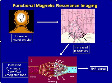
A person has a thought or idea or does some cognitive or perceptual task this leads to an increase in neural activity – in a specific region or regions of the brain – a FOCAL increase in neural activity.
This in turn leads to a FOCAL increased blood flow to that same brain region which leads to increased oxygen delivery to that region – (or more accurately a change in the ratio of deoxy and oxy hemoglobin).
It is this change in oxygen that gives us our signal in fMRI
So one thing to note from this is that this signal that is measured – the change in oxygen – is produced naturally by the body - no contrast needs to be injected. This means it is a non invasive procedure, unlike other forms of brain imaging like PET scanning which requires an injection of radioactive materials. This means fMRI can safely be used to scan children’s brains and it can also be performed multiple times in the same person – which means it is possible to now look at the effects of training, interventions, etc.
The second thing to note from this is that this signal measured – while it is inferred to be representative of neuronal activity – is an INDIRECT measure of brain activity. That is an important limitation to remember about the method – at this point the primary way of measuring neuronal activity directly is still limited to animal studies.
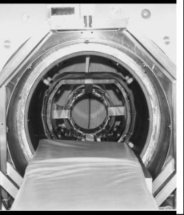
An Ultra-fast fMRI Scanner (field strength: 2 T), Hitachi Central Research Laboratory, 1992
Copyright Hideaki Koizumi
See also:
-
fMRI Beyond the Clinic: Will It Ever Be Ready for Prime Time? PLOS Biology Vol 2, Issue 6, June 2004.
Related Documents